Implants: “You don’t know what you can’t see”
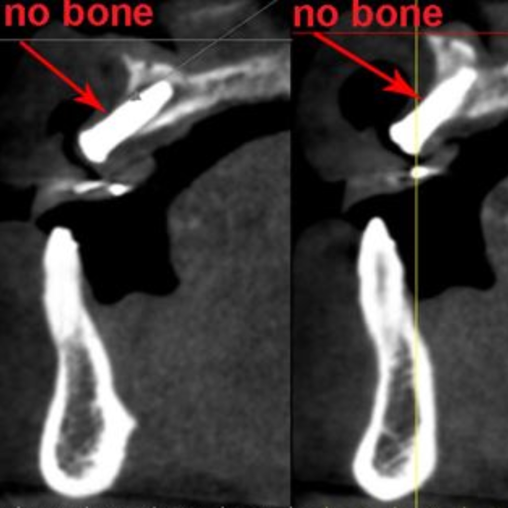
3D dimensional imaging has become an important tool in our office. In approximately 8 secondss we can get a full 3D view of the upper and lower jaws and adjoining sinuses. The images or slices can be viewed several different ways. Picture an apple that is sliced many times from top to bottom (the long way). The apple slices can then be viewed from what is called a cross sectional viewpoint. Supposing the apple is sliced many times parallel to its equator (the short way). The slices can then be viewed from what is called an axial viewpoint. We can also view the apple as a normal unsliced 3D object.
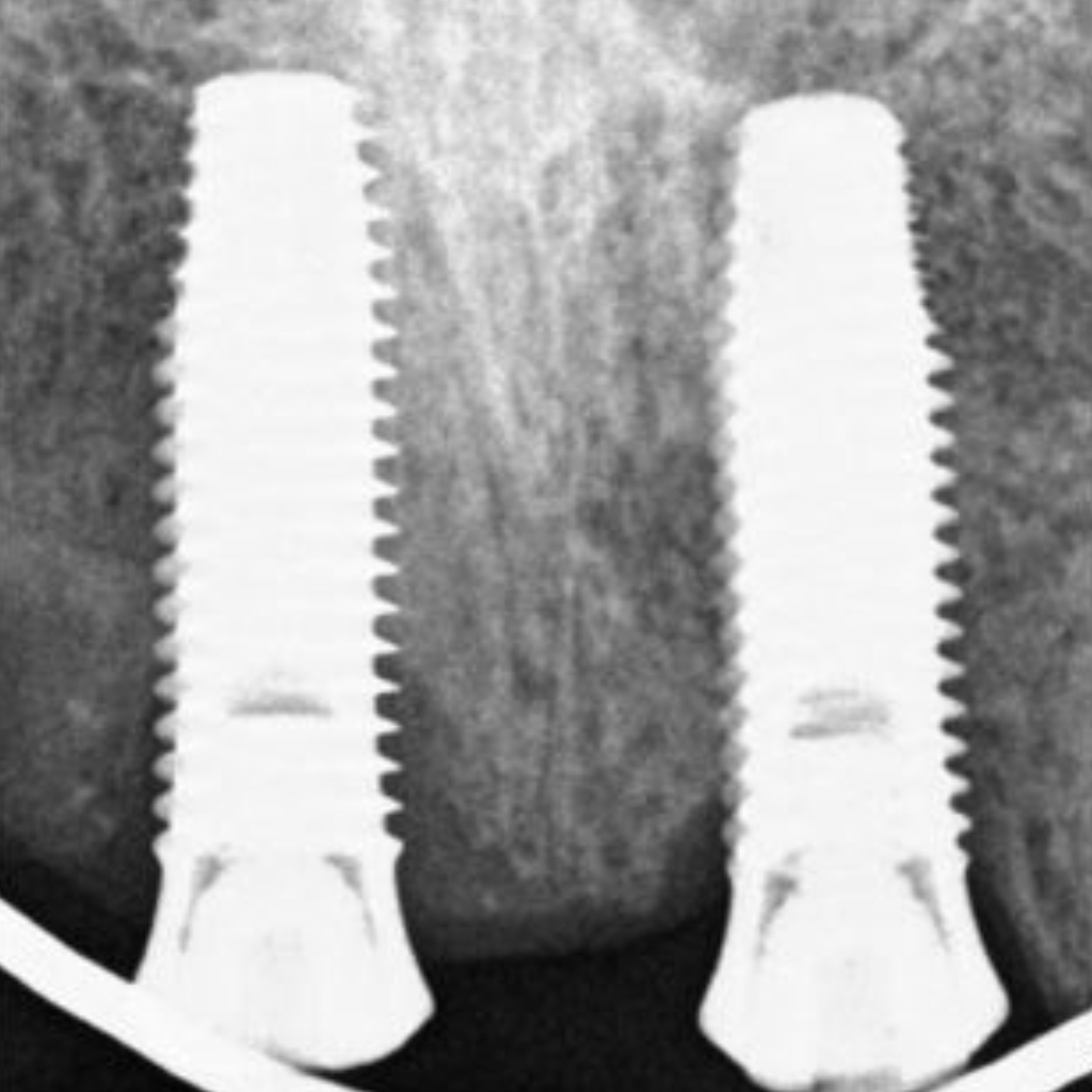
A regular 2 dimensional x-ray does not nearly give us the same information and can often times be misleading. In the x-ray below we see two implants that appear to be fully in bone. There is basically nothing to concern us.